Mitosis In Animal Cell Whitefish Blastula / Whitefish Blastula Mitosis 320x Stock Photo Picture And Rights Managed Image Pic Aam Aaes80601 Agefotostock - Whitefish blastula (animal cell mitosis)
byKendra Ronhaar-
0
Mitosis In Animal Cell Whitefish Blastula / Whitefish Blastula Mitosis 320x Stock Photo Picture And Rights Managed Image Pic Aam Aaes80601 Agefotostock - Whitefish blastula (animal cell mitosis). Stages of the cell cycle in animal cells (whitefish blastula) cell cycle stage sketch interphase prophase metaphase anaphase telophase During this phase all cells are actively dividing and growth is rapid. Two specimens are commonly used by biologists to study mitosis: Click here to begin the tutorial Examine prepared slides of both the onion root tips and whitefish blastulas.
Examine all sections on the slide using high power to locate all the phases of mitosis. About press copyright contact us creators advertise developers terms privacy policy & safety how youtube works test new features press copyright contact us creators. | certified educator there are many similarities between mitosis in plant cells, such as onion cells, and animal cells, such as whitefish blastula. These cells are constantly dividing cells just like meristematic cells of plants. Focus on one that is in good condition and in which the cells are easy to see.
Cell Cycle And Mitosis Laboratory Notes For Bio 1003 from faculty.baruch.cuny.edu The mitotic stages are clearly visible in these cells. Key characteristics of animal cell cytokinesis: Spindle and aster are visible. Determining the rate of mitosis in plant and animal cells. Looking at 2 or more. You will make observational drawings and be prepared to take a practical quiz. Whitefish blastula (animal cell mitosis) Observation 6 animal cell mitosis obtain the slide of a whitefish blastula.
Observation 6 animal cell mitosis obtain the slide of a whitefish blastula.
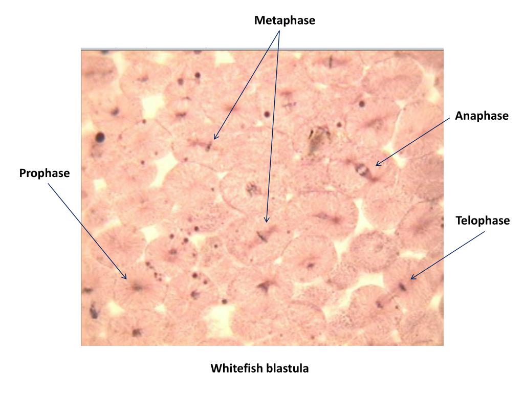
Practice locating each of the stages of mitosis in the following photomicrographs. Examine prepared slides of either onion root tips or whitefish blastula. So, for studying mitosis in animal cells, generally whitefish blastulas are used. Whitefish chromosomes are small so detailed observation of chromosome structure will be difficult, however, the spindle fibers of the mitotic apparatus are very beautiful. Study the successive stages of mitosis in the cells of the white fish blastula. Whitefish blastula cells are used for studying mitosis. Whitefish blastula and onion root tip are selected for the study of mitosis because these two tissues have many cells that are in the process of mitosis. Mitosis in plant cells (onion root tip) mitosis in animal cells (whitefish blastula) · · · • • • • • · · · The human chromosomes will not be clearly. Über 7 millionen englischsprachige bücher. Stages of the cell cycle in animal cells (whitefish blastula) cell cycle stage sketch interphase prophase metaphase anaphase telophase There are very few similarities between animal cell and plant cell cytokinesis. Figure 3.1 close up view of different stages of mitosis in an onion root tip:
These haploid cells generally become gametes. Please note that if you will use human blastula instead of whitefish blastula; Key characteristics of animal cell cytokinesis: Spindle and aster are visible. Click here to begin the tutorial
Ppt Whitefish Blastula Powerpoint Presentation Free Download Id 2373110 from image1.slideserve.com The human chromosomes will not be clearly. The developing embryo of any organism is a good tissue to examine for mitosis, since cells must divide at a high rate to transform a fertilized egg (single cell) into the trillions of cells of a viable organism. These cells are constantly dividing cells just like meristematic cells of plants. Find each phase of mitosis on the slide. Mitosis in whitefish blastula cells: Mitotic activity in animals is most rapid during early development. You may use your textbook and class notes to help you identify the stages of mitosis as seen under the microscope. In the photograph the arrow points to a cell in anaphase.
There are very few similarities between animal cell and plant cell cytokinesis.
Study the successive stages of mitosis in the cells of the white fish blastula. The blastula is an early phase of development found in animal embryos; The human chromosomes will not be clearly. Mitosis in animal cells whitefish (leucicthys) blastula slides examine the prepared slide of whitefish blastula in your tray. The blastula of a whitefish and the root tip of an onion. As you locate each phase, your instructor will verify that you are correct and initial your paper. There are several cross section cuts on each slide; Note that sections of six different blastulae are mounted on this slide. Mitotic activity in animals is most rapid during early development. Focus on one that is in good condition and in which the cells are easy to see. I focused the microscope on the 4x setting with the onion root tip in view, making sure the diaphragm again was at its widest setting. Whole mounts of whitefish blastula will illustrate reproductive cells in animals. In this lab, you will determine the approximate time it takes for a cell to pass through each of the four stages of mitosis.
Mitosis in animal cells (whitefish blastula) meiosis meiosis is a type of cell division that begins with a single diploid cell and finishes with four haploid cells. Therefore, the blastula stage of a whitefish embryo has been selected for observation. Early in interphase the cell (a) reaches its full size and then starts preparing for its next division. The mitotic stages are clearly visible in these cells. Note that sections of six different blastulae are mounted on this slide.
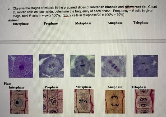
B Observe The Stages Of Mitosis In The Prepared Chegg Com from media.cheggcdn.com First, scan the slide using low power. I focused the microscope on the 4x setting with the onion root tip in view, making sure the diaphragm again was at its widest setting. Looking at 2 or more. Observation 6 animal cell mitosis obtain the slide of a whitefish blastula. The blastula of a whitefish and the root tip of an onion. Early in interphase the cell (a) reaches its full size and then starts preparing for its next division. The blastula is an early stage of embryo development, and represents a period in the organism's life when most of the cells are dividing consistently. Observing mitosis in plant and animal cells using prepared slides of the onion root tip and whitefish blastula.
Determining the rate of mitosis in plant and animal cells.
These haploid cells generally become gametes. The cell will arrest in mitosis and die.4. You will study the crossing over and recombination that occurs during meiosis. There are several cross section cuts on each slide; Spindle and aster are visible. The four steps of mitosis are prophase,. Whitefish mitosis practice the photomicrographs below show sections of whitefish blastula. In this lab, you will determine the approximate time it takes for a cell to pass through each of the four stages of mitosis. Figure 3.1 close up view of different stages of mitosis in an onion root tip: First, scan the slide using low power. In animal cells, a ring of actin fibers is formed around the periphery of the cell at the former metaphase plate (cleavage furrow). You may use your textbook and class notes to help you identify the stages of mitosis as seen under the microscope. The blastula is an early phase of development found in animal embryos;